A new home for our Mass Spec prototype
Our prototype stigmatic imaging secondary ion mass spectrometry (SIMS) instrument has been moved from development laboratories at Ionoptika’s factory to the Department of Chemistry at the University of Oxford. This exciting development reflects the progression of this project to its next stage.
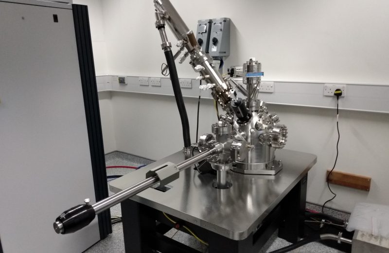
The stigmatic imaging SIMS instrument will enable rapid molecular mapping of biological tissues at unprecedented speed, as this type of mass spectrometry imaging decouples acquisition time from spatial resolution. Typically mass spectrometry imaging scans across a surface taking a mass spectrum at each spot to build up the pixels of the image.
In this case, however, the whole surface is imaged simultaneously using state of the art cameras that operate as an array of position and time sensitive detectors that record a mass spectrum for each pixel in the camera image. It therefore represents a promising route to attaining higher throughput.
This instrument design is a joint endeavor between researchers at The Franklin, the University of Oxford and Ionoptika. Ionoptika is an ion beam technology company with a team of scientists and engineers that are committed to the development of high performance ion beam technology for scientific analysis applications such as secondary ion mass spectrometry and ion implantation. The first stage of this project took place at Ionoptika’s headquarters near Southampton, where this unique instrument was built and tested.
Dr Felicia Green, part of the Mass Spectrometry team at the Franklin and a key part of the instrument development team, said, “I am thrilled to be part of this project, it really has the potential to greatly improve mass spectrometry imaging.”
Dr Green added: “Prior to moving the instrument, I was able to spend time at Ionoptika headquarters, testing the instrument. This was a great experience and the team at Ionoptika were brilliant and very efficient. I am looking forward to continuing to work with the team at Ionoptika and the University of Oxford to continue to develop this prototype.”
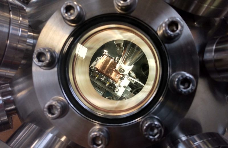
Now that the instrument has been installed at the University of Oxford, initial characterization and alignment of the primary ion beam will first need to be carried out, before the addition of extraction optics and high speed stigmatic detectors. With these further developments and improvements to this instrument, we hope to achieve a step change in microscope mode resolutions while maintaining a broad mass range capability. This would mean a mass spectrometry image of a standard tissue biopsy would take seconds rather than hours or days, paving the way for routine analysis.
Information gathered from this prototype will be used to build the next level instrument. This next level instrument will be built at the Franklin, either as part of, or complimentarily to, the larger Biological Mass Spectrometry instrumentation.
Read more about this instrument or the broader Franklin Biological Mass Spectrometry theme here.