New cutting-edge microscope for the UK
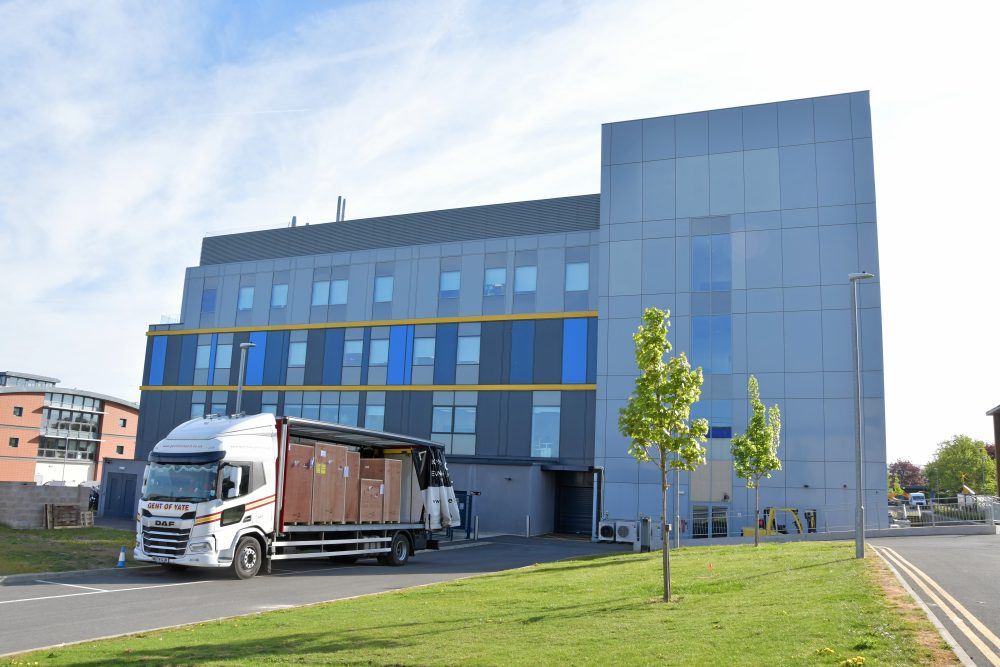
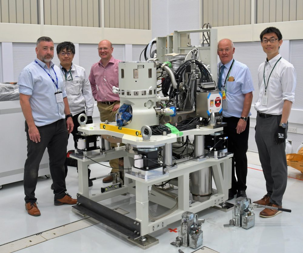
The UK’s first Chromatic Aberration-Corrected Electron Microscope has arrived at the Rosalind Franklin Institute. This state-of-the-art instrument will significantly enhance the resolution limits for biological sample imaging, especially for thicker specimens.
This delivery marks the final instalment of three advanced instruments developed in collaboration with JEOL Ltd, a leading electron microscope manufacturer, headquartered in Japan.
Mr. Shaun Quill, Managing Director of JEOL (UK) Ltd. said, “The longstanding collaboration between the Franklin and JEOL has reached a noteworthy milestone with the delivery of this newly developed, state-of-the-art instrument. This achievement is the pinnacle of several years of dedicated joint effort, during which both parties have worked closely to realize this significant advancement in electron microscopy.
We are proud to announce that the Franklin is not only the first institution in the United Kingdom but also the first globally to receive JEOL’s pioneering Chromatic Aberration-Corrected Electron Microscope.
This instrument will complement the existing suite of JEOL technologies at the Franklin, collectively representing the forefront of electron microscopy and advancing the capabilities of life science research to unprecedented levels.”
The unique suite of electron microscopes is enabling researchers at the Franklin to advance new techniques, especially in the field of liquid phase electron microscopy. Liquid phase microscopy allows researchers to study molecules and larger biological structures in motion. When studying the molecules of life, the ability to view them in their ‘native’ environment, and watch their interactions in real-time will be critical.
Franklin researchers have already made significant progress in this field imaging bacteria in liquid with unprecedented clarity. This progress was possible to advancement in graphene cell technology developed at the Franklin by Dr Adrian Pedrazo-Tardajos.
The new microscope will improve the resolution limits for biological samples even further, as its chromatic aberration correction will correct for energy variations in the electron beam. These limit resolution, particularly for thicker samples such as liquid cells which often use Silicon Nitride (SiN) membranes to encapsulate films of liquid.
Professor Angus Kirkland, Science Director at the Rosalind Franklin Institute, said, “We are very excited by the arrival of this unique instrument which represents the culmination of a long-standing collaboration between the Franklin and JEOL. We are confident that this will enable us to push the boundaries of imaging, with the long-term goal to image biological structures in their native environment and to observe their dynamics in situ.”
Read more about Electron Microscopy Platforms at the Franklin.