Major step in our cellular tomography project
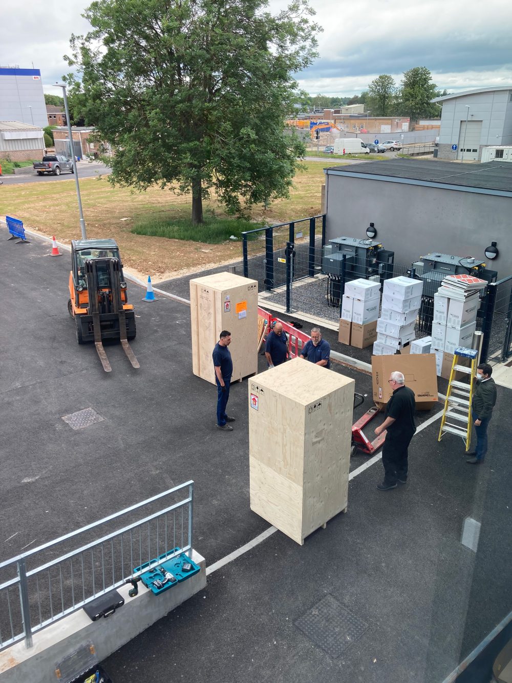
The Rosalind Franklin Institute has taken delivery of its first Titan Krios microscope, named Dorothy, and the second generation of the plasma focused ion beam (pFIB), named Franklin, both from Thermo Fisher Scientific. These new technologies will utilise new sample geometries to enable high resolution, large volume cellular tomography for the first time.
Large volume cellular tomography has the potential to transform our understanding of life, enabling us to view proteins to atomic resolution within the cell. Utilising the unique capabilities of these technologies will allow researchers to address more complex biological questions, including deepening our understanding of intracellular bacterial pathogen life cycles. Intracellular bacterial pathogens are becoming increasingly resistant to existing antibiotics, so the ability to observe the bacteria life cycle inside the human cell could be a critical first step in developing new drugs to combat it.
The large volume cellular tomography technology is being co-developed by scientists and engineers at the Rosalind Franklin Institute, Thermo Fisher Scientific and Diamond Light Source, this project is funded by Wellcome through the Electrifying Life Science grant.
Steve Reyntjens, Product Marketing Director for Thermo Fisher Scientific said, “Thermo Fisher is a company driven by the needs of scientists. It has been thrilling to work with the Franklin because they want to make breakthrough innovations, not just incremental changes, so working to meet their scientific needs has been a real challenge.”
Critical innovations are required for this project to be successful, not just in the design of the instrument, but in the data collection and processing software which is used. Powerful data processing is needed to manage the volume and type of data produced, so close collaboration with The Franklin Artificial Intelligence and Informatics theme has proven to be essential.
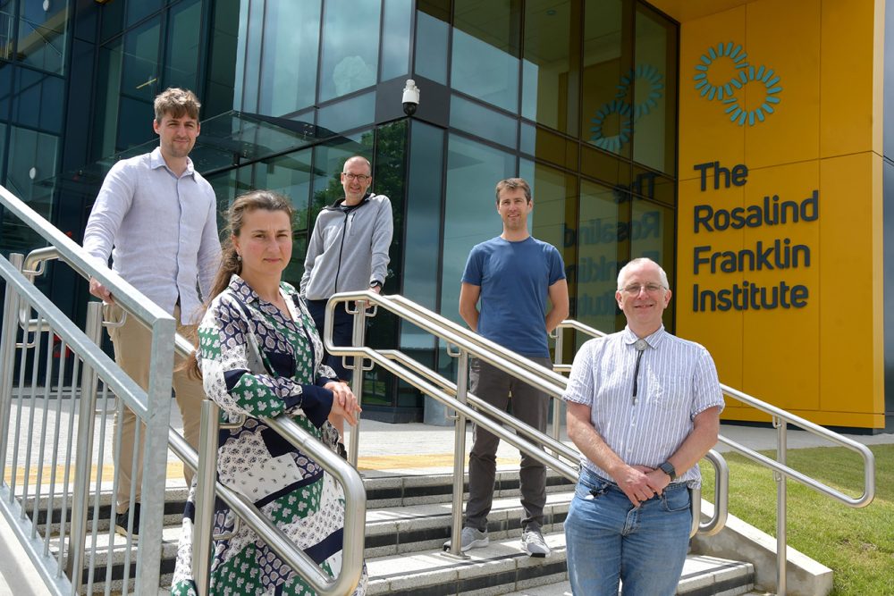
Dr Maud Dumoux, The Franklin’s Technology Lead for Cryo Imaging, said, “The project is a challenging and exciting to be a part of; it has the potential to revolutionise how we see the cell. Improving our understanding of this basic building block of life could have a profound impact on how we create new drugs and understand how they work.”
Plans are already in motion for the next generation KRIOS microscope to be built as a collaboration between Thermo Fisher Scientific and the Franklin, which is set to be delivered in 2024. There is already a clear vision for this machine, which will contain a special aberration corrector to allow more information to be collected more quickly from sample. The knowledge gained from Dorothy will be crucial to inform the design of this beyond state-of-the-art machine, which will be known as Hodgkin.
Professor James Naismith, Director of the Franklin and Science Director for Structural Biology, said, “The delivery of Dorothy marks the start to the next stage of this project and we are all excited to see exactly to help usher in the era of atomic cell biology. The Franklin are excited to be part of this and committed to sharing our new advances in technology and approaches with the widest community.”