New Grant will Electrify Life Sciences with Breakthrough Technologies
The Rosalind Franklin Institute, working with partners MRC Laboratory of Molecular Biology (MRC LMB) and Diamond Light Source, have been awarded a £25m grant from Wellcome to support the development of a trio of electron imaging physical sciences technologies with the capacity to revolutionise how we see life. The overall project will be led by Professor James Naismith, Director of the Franklin, Professor David Stuart, Director of Life Sciences at Diamond Light Source, and Dr Richard Henderson, group leader at the MRC LMB.
Collectively known as ‘Electrifying Life Science’, the electron imaging technologies will create globally unique capabilities for the UK. Together, the team will change by a factor of ten the accessibility and capability of electron cryomicroscopy (cryo-EM), in both tomography and single particle sub-fields.
The Amplus project, led by the Franklin, will deliver a revolution in cryo-electron tomography – using electron microscopy to build up three dimensional models inside the cell. This will bring into view the possibility of imaging whole cells at atomic scale quickly and accurately. There are huge technical challenges to overcome in cryo-electron tomography, from sample milling, handling, and imaging, plus data reconstruction. However, the pay-off from accomplishing these technical challenges will be enormous, with applications in genetic disease, intracellular pathogens, understanding the mechanisms of microbial drug resistance, and observing viral infection.
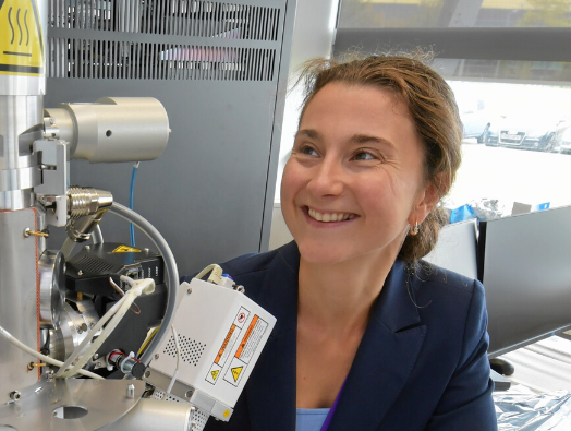
Dr Maud Dumoux, the Franklin’s postdoctoral scientist working on Amplus, said, “The Amplus project is a challenging and exciting project to be part of, it has the potential to revolutionise how we see the cell. Improving our understanding of this basic building block of life could have a really meaningful impact on how we create new drugs and understand how they work within the cell.”
To make the process robust and accessible, automation will be built in to every step, from sample handling to data processing. Amplus will be built at the Franklin Hub, with beyond state-of-the-art microscopes and associated equipment.
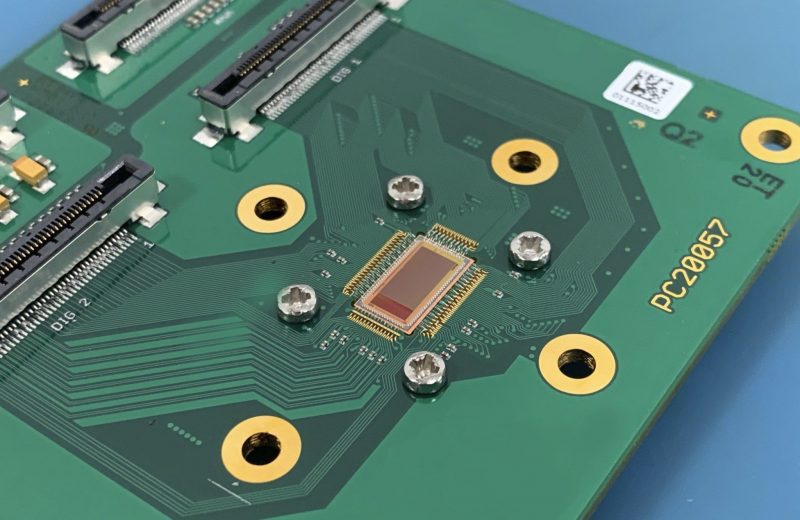
Technology developed with MRC LMB will democratise access to electron cryomicroscopy. Working with the technique’s pioneer and Nobel Prize winner, Dr Richard Henderson, and LMB group leader Dr Chris Russo, the team will develop instrumentation to enable single particle cryo-EM at 100 keV, as opposed to industry standard 200 and 300 keV machines with no loss of quality in resolution. This reduction in energy drives down the cost of equipment, and reduces the environmental requirements of the microscopes, taking the technique out of highly specialist spaces and into everyday laboratories. This democratisation will enable specialist labs to undertake more complex work, raising the standards of cryo-EM across the board.
Dr Jan Löwe, Director of the MRC LMB, said, “The LMB has helped to pioneer the development of biological applications of electron microscopy. Through the exciting and vast collaboration bringing together institutes, facilities and commercial instrumentation specialists the method will become more accessible, faster, more precise and more generally applicable. It will help to make electrons not only the best but also the preferred option to investigate, and to utilise, the secrets of life.”
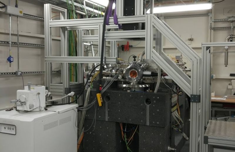
Another technology development led by Professor Stuart and Dr Gwyndaf Evans, a Diamond and Franklin group leader, will be based at Diamond Light Source, the UK’s national synchrotron. The Hybrid electron – X-ray Instrument, known as HeXI, will play a major role in drug discovery efforts and when combined with X-ray methods will enable crystallography on small crystals ranging from around 100 nm, approximately 100 times thinner than a human hair, to up to 10 µm (ten millionths of a metre).
This technology will be a dedicated electron diffraction instrument embedded within Diamond’s VMXm micro/nano focus X-ray beamline facility. Upon completion HeXI will be accessible through the Diamond user programme.
“HeXI will combine and build upon state-of-the-art technologies from electron and X-ray fields to create brand new scientific opportunities for structural biology and drug discovery. It will make electron diffraction easily accessible to all of Diamond’s existing life science users and attract new users to routinely study pharmaceutical compounds and their binding,” commented Dr Gwyndaf Evans, a Diamond and Franklin group leader.
This grant will also allow training in all of these advanced techniques to both UK and international scientists.
As a suite of technologies, the Electrifying Life Science grant will push the boundaries of the current capability of imaging in life science, while opening up the technique to a wider audience than ever before, and creating technological links with existing techniques.
Professor James Naismith said, “This high-risk, high-reward multidisciplinary project is what the UK Government set up the Rosalind Franklin Institute to do. I cannot emphasise enough the team work required to get us to this stage and how important it will be going forward. Electrifying Life Science will be a true factor of ten change in our ability to see and understand life. We are incredibly grateful to Wellcome for placing their trust in us. I want to acknowledge funding from UKRI-EPSRC, core funding partners of the Franklin, and of course from our industrial partners who are contributing their world leading engineering and design capability. Everyone at the Franklin feels humbled by this and are excited to start work to deliver on our promises.”
Dr Tom Collins, interim Head of Genetics & Molecular Sciences at Wellcome, said “There has been an explosion in the scientific impact of cryo-electron microscopy since the introduction of revolutionary electron detector technology. We are excited about this project that gives us an opportunity to bring closer together structural and cell biology through the development of the next generation of this innovative technology.”