
Dr Brian Caffrey

Currently, Brian is working in collaboration with the Rosalind Franklin Institute and the University of East Anglia developing novel imaging methods which combine liquid-phase, conventional cryo-electron microscopy, spectroscopy, and correlative light microscopy techniques to study biological processes in situ. In particular, his research focuses on applying these techniques to study antimicrobial resistance mechanisms to aid in the development of novel antimicrobials.
Brian graduated with a BSc in Chemistry from University College Dublin (IRL). During his time at UCD he also gained industry experience working on pharmaceutical chemical engineering projects at APC Ltd. (IRL). These formative experiences fuelled his interest in the molecular basis of disease and in creating tools to better understand and treat it.
He was awarded a joint Wellcome Trust / National Institutes of Health PhD studentship, completing his doctorate in Biochemistry and Molecular Biology between the National Cancer Institute (USA) and the University of British Columbia (CA) in Prof. Sriram Subramaniam’s lab. His PhD focused on developing 3D electron microscopy methods to interrogate molecular and organellar structures involved in cancer, aging and neurodegenerative disease. Notably, his cryo-EM work on the p97 ATPase revealed mechanisms of allosteric inhibition and supported the development of therapeutic candidates that have progressed to preclinical testing. At the same time, he also taught as Adjunct Professor at Georgetown University in the School of Nursing.
After completing his PhD, Brian worked at Gandeeva Therapeutics (CA), applying cryo-EM structure-aided drug design and developing mammalian cell-based assays for screening therapeutics before moving to a postdoctoral position at the Rosalind Franklin Institute where he developed liquid phase electron microscopy methods for structural biology applications.
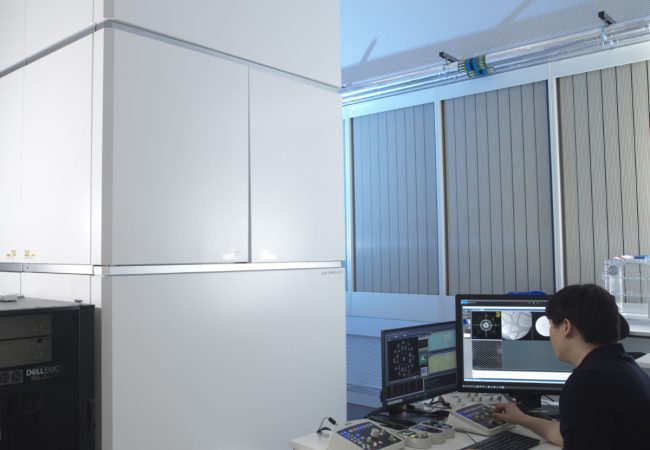
Aberration-corrected transmission electron microscope
Ruska is an aberration-corrected transmission electron microscope (TEM) used to explore novel methods to study radiation sensitive specimens such as biological materials that have been cryogenically preserved or encapsulated in liquid for dynamic observations.
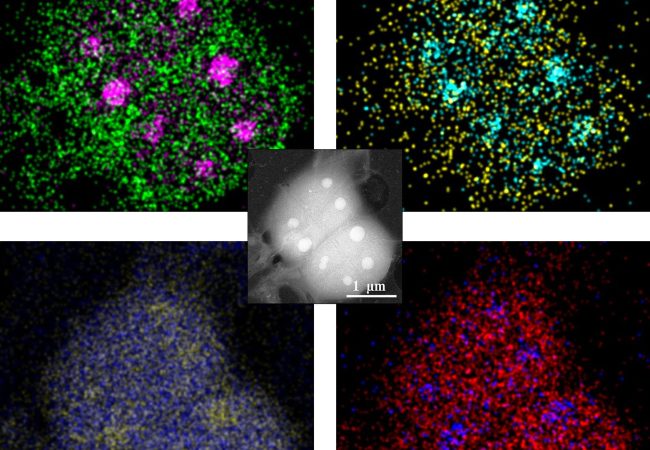
Liquid Phase Electron Microscopy and Spectroscopy
Transient, dynamic assemblies of biomolecules in solution are the primary driving forces behind biology. However, studying these at high resolutions requires the use of electron microscopes (EM), which need extremely high vacuums to function.