Mass Spectrometry Imaging
We are developing an array of instrumentation mass spectrometry instruments that will improve the resolution, accuracy and sensitivity of mass spectrometry imaging of these instruments, so that we are able to analyse and interpret complex cellular and molecular information.
Through these developments of this instrumentation, we are looking for ways to unlock the most information possible from our biological samples across scales from large protein assemblies to small metabolites.
Instruments
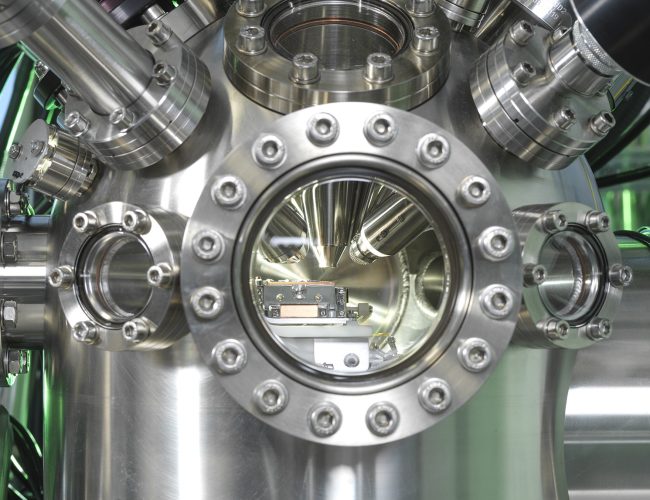
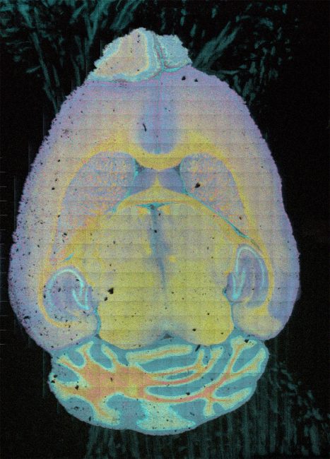
High Resolution imaging with secondary ion mass spectrometry (SIMS)
Secondary Ion Mass Spectrometry (SIMS) is a highly sensitive analytical technique offering detailed chemical composition analysis in 3D space with subcellular resolution.
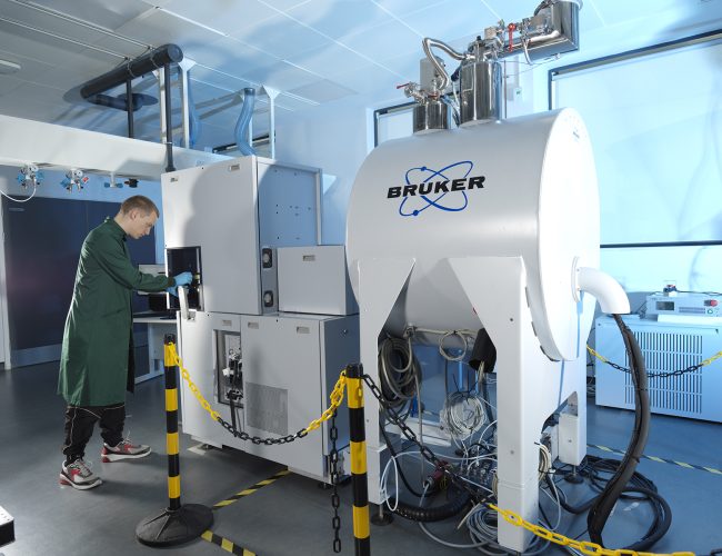
Trapped ion mobility (TIMS) time of flight (TOF) mass spectrometry
A cutting-edge commercial Bruker mass spectrometry (MS) instrument, coupling high sensitivity, high resolution, rapid time of flight (TOF) mass analysis to high resolution trapped ion mobility spectrometry (TIMS) enabling structural elucidation.
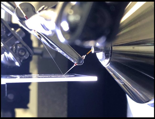
Native ambient mass spectrometry
Native ambient mass spectrometry (NAMS) combines spatial and structural biology by enabling untargeted label-free interrogation of proteins in their functional form directly from their physiological environment.