Unique microscope to film molecules in motion
A unique electron microscope, set to break new ground in biological imaging, will arrive at the Franklin’s new building on the Harwell campus today. The machine is able to image biological samples at up to a million frames a second, a thousand times faster than the current standard.
It is the first of three instruments being developed jointly with electron microscope manufacturer, JEOL Ltd, shipped to the UK from the company’s headquarters in Japan.
‘Ruska’ will work with biological samples both cryogenically frozen and in liquids, which will enable imaging of molecules in motion. By combining high speeds with liquid cells, the Ruska microscope will be able to ‘film’ proteins as they fold or image drugs interacting with other molecules. For cryogenically frozen samples, the frames captured as the beam passes through the sample will enable the creation of 3D models of biological structures, such as viruses or proteins.
The Correlated Imaging Team at The Franklin have adapted an approach widely used in physical sciences for use in biological imaging. The technique provides images at higher contrast than traditional TEM imaging, but is more commonly used with materials such as semi-conductors or catalysts.
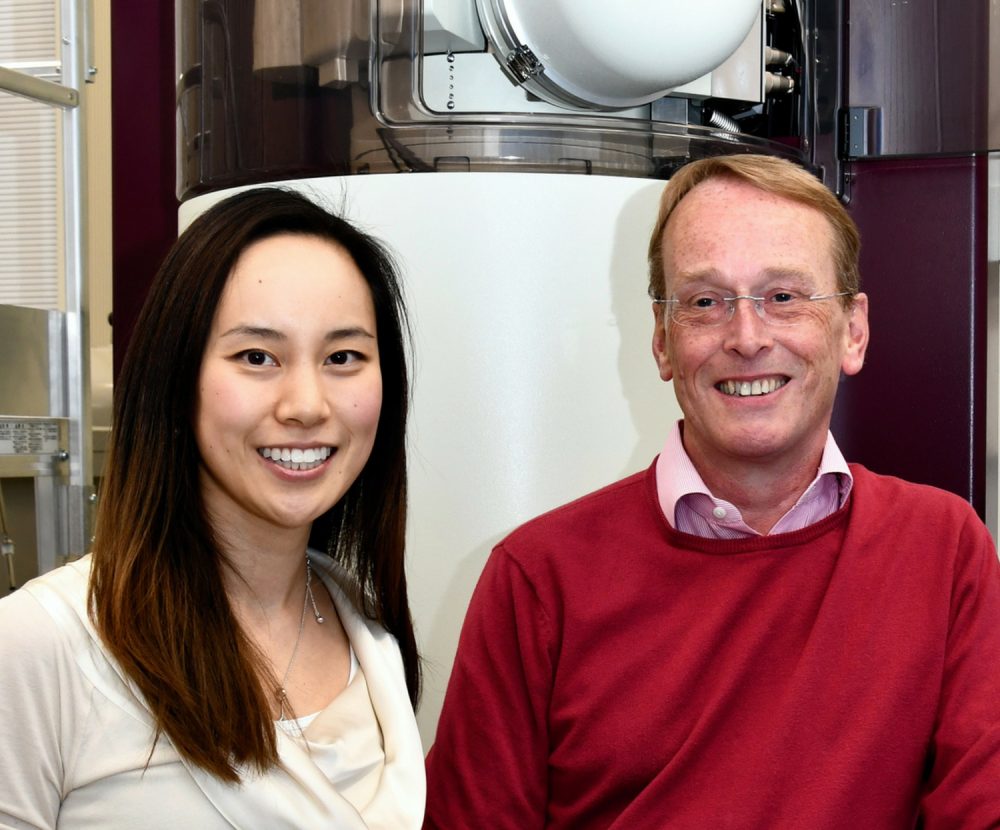
Correlated Imaging Deputy Director, Dr Judy Kim said: “We’ve already shown in initial experiments that we can use this technique to look at biological material. But the machine we were using wasn’t optimised for these kinds of samples. With the new machines, we’ll be able to refine the technique further. We’ll also be able to run very fast, to record data at close to a million frames a second, which will reduce the radiation damage visible on the sample.”
The machines are among the first equipment to be installed in the Franklin’s new building on the Harwell Campus, which opens fully later this year.
A specialist area has been created for the electron microscopes which minimises vibration, magnetic fields and acoustic noise and is also very carefully temperature controlled.
Correlated Imaging Director, Professor Angus Kirkland explained: “We’re looking at features that are smaller than the wavelength of normal visible light, so even tiny vibrations or changes in temperature can cause things to move. The machines have to be in a tightly controlled environment, so when we operate them, which we do remotely, there’s no risk of environmental interference.”
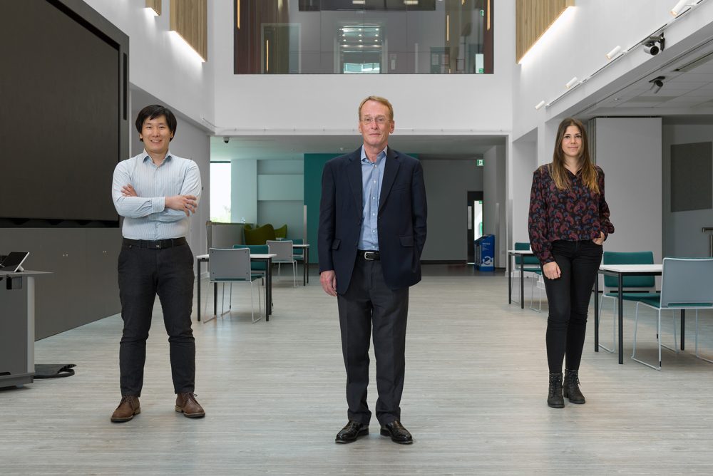
It will take several months of work to fully commission and calibrate the instruments once they are installed in their new home. A team of four will work full-time with the microscopes, including researchers from both physical science and biological sciences backgrounds. In addition, researchers based at the Franklin’s various partner institutions will continue to work on specific aspects of the new technology and processes, such as liquid cells.
For Professor Kirkland and Dr Kim, the arrival of the machines brings both a sense of relief and anticipation.
Dr Kim said: “Until now, we’ve been planning so much, so to have it all come together and see the machines arrive is really wonderful. We know these instruments will work, so we just have to find out to what level. How fast will we be able to go, or to what resolution? It really is very exciting.”
Professor Kirkland agreed: “The last five years have been very much about designing the instruments and building the platforms. But the next five years is about using those to do some really exciting science. We can’t wait to get started.”
Short animation explaining our new microscope, Ruska: