Using mass spectrometry imaging to understand metabolic mechanisms of disease
Working with industry and academic partners, we are developing secondary ion mass spectrometry (SIMS) as a technique to study highly detailed biomolecular information at cellular level. This is allowing us to study cellular health, from metabolism to energetics, in greater detail and resolution than before.
This instrumentation is now fully operational at the Franklin and is already being used by researchers in the UK and internationally to understand how disease begins and progresses. This includes research to understand the early biological changes in Alzheimer’s disease and motor neurone disease, and in an international piece of research to understand cell death in psoriasis.
Identifying the biological changes that lead to disease will give the foundations to develop pioneering new treatments in future.

The instrument developed in collaboration with UK based Ionoptika enables a dramatic improvement in mass spectrometry imaging (MSI). The new instrument is capable of imaging the whole surface simultaneously, allowing more accurate imaging at faster speeds than was previously possible.
Background
Working with industry and academic partners, we are developing secondary ion mass spectrometry (SIMS) as a technique to study highly detailed biomolecular information at cellular level. This is allowing us to study cellular metabolism and energetics in greater detail and resolution than before.
This instrumentation is now fully operational at the Franklin and is already being used by researchers in the UK and internationally to understand how disease begins and progresses. This includes research to understand the early biological changes in Alzheimer’s disease and motor neurone disease, and in an international piece of research to understand cell death in psoriasis.
Identifying the biological changes that lead to disease will give the foundations to develop pioneering new treatments in future.
Science in detail
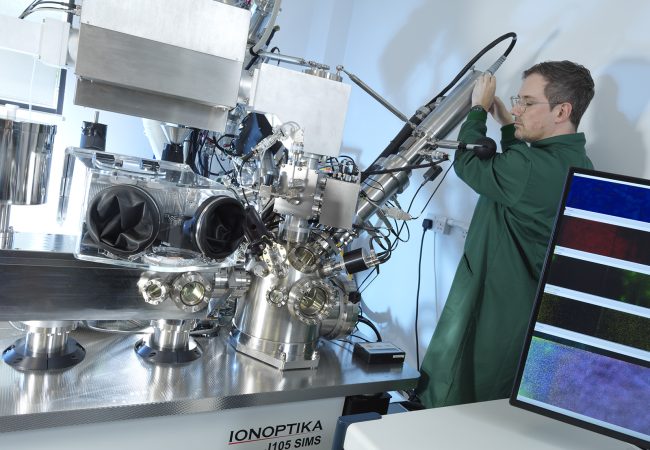
To make this possible, the Franklin worked with industry partner Ionoptika, and collaborators at the University of Manchester, using a world-leading secondary ion mass spectrometry instrument that is equipped with a water cluster ion beam and a capability to image frozen samples. This instrument can collect highly detailed biomolecular detail from cryopreserved samples at cellular resolution.
To do this a water cluster ion beam gun is used that allows considerably more chemical information to be gained from a sample than is possible with more conventional approaches. The instrument that was developed also allows tissue samples that have been cryopreserved to be analysed. This allows the study of biological processes that have been stopped in time to be studied with reduced artifacts from pathological processes that occur when tissues are warmed outside of the body.
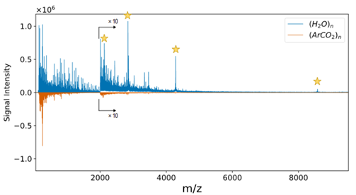
The 70keV ion beam produces clusters of water molecules with as many as 50,000 molecules per cluster. These are fired at high acceleration onto the surface of the sample but break apart on impact, where they gently desorb molecules from the surface such that they then can enter the mass spectrometer. The introduction of large water gas cluster ion beams (GCIB) reduced undesirable molecular fragmentation and enhanced ionization efficiency due to the large size and proton-rich nature of the primary ions, respectively. The Franklin scientists then showed that, compared to (Ar/CO2)n clusters, water clusters produce substantially higher ion yields for peptides and small proteins. This translates to improved sensitivity. Notably, water clusters facilitated the generation of multiply charged species, including, for example, the detection of 3+ and 4+ charge states for ubiquitin. This ability to form and detect multiply-charged protein species represents a major advancement for SIMS analysis, because it promises the opportunity to expand its applicable mass range to large proteins and biomolecules.
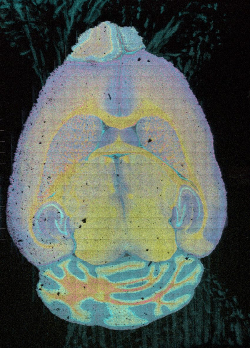
This is one of the few SIMS instruments worldwide at this time that is equipped with a water cluster ion beam for analyses of cryopreserved biological samples. It allows researchers to analyse cells and tissues at a cellular scale to get a wealth of biomolecular information about the chemical, energetic and metabolic status of cells and tissues in three dimensions. This includes the full lipidomic and metabolomic profiles.
This biomolecular technique is being applied to another advance in imaging: the integrated imaging of cell chemistry and structure: cryo preserved samples suitable for subsequent SEM and TEM analysis can be first analysed for their biomolecular composition. The emerging impact has been recognised by additional funding to explore the transfer and suitability for multi-dimensional imaging between analytical techniques.
Another area of Franklin research that has benefited from the new kit, is a project to understand how Chlamydia trachomatis LGV2 serovar infects cells. The technique is allowing researchers to track the lipidomic changes as bacteria infect cells, enabling an understanding of the biomolecular differences between infected and uninfected cells.
The new instrument has also been used by researchers outside the Franklin. A local SME, called NeuroBio, used the kit in their research to understand how new early biomarkers of Alzheimer’s disease affect the early mechanisms of neurodegeneration. In addition, a visiting scientist from University of Pennsylvania has used the instrument to understand a process called ferroptosis, a form of iron-dependent cell death, which seems to play an important role in the development and progression of psoriasis.
We are continuing to develop capabilities for this new instrument, and sharing our expertise in water cluster ion SIMS with the wider scientific community in the UK and internationally. By interacting with potential new users we have helped the company, Ionoptika Ltd based in Southampton, to sell 2 new instruments that will be delivered in 2025/2026.
Why the Franklin?
The Franklin has proved to be a uniquely powerful environment for development of this work. The project involved interdisciplinary interactions between mass spectrometry and biological applications experts. It benefited from analytics capabilities of the Franklin’s ARC and AI teams. Finally, the future road map for integration of mass spectrometry and ultrastructural imaging is being driven by the colocalisation of collaborating mass spectrometry and EM teams.