Developments in cryo-electron ptychography
Researchers at the Rosalind Franklin Institute working alongside colleagues at Nanjing University, University of Warwick, Diamond Light Source and University of Oxford are adapting an electron microscopy technique for life sciences which was originally developed for the materials science. Using this technique, they have achieved <2nm resolution of Rotavirus particles.
Their findings have recently been published in Nature Communications.
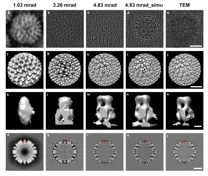
The technique, known as cryo-electron ptychography, allows scientists to gain high resolution images with enhanced contrast. The technique differs from other electron microscopy techniques as it uses a defocused electron beam to scan across a sample. By computationally reconstructing the images from 4D diffraction datasets, the team are able to collect more information about the same point in the sample which allows them to combine low and high spatial frequency data.
There are a number of advantages of this technique over standard transmission electron microscopy and scanning transmission electron microscopy, in particular fewer particles are required to make a high-quality 3D reconstruction. This is useful for highly heterogeneous biological samples.
It also allows researchers to produce high resolution images with improved contrast and dose efficiency, which is especially important for biological samples as they tend to be quickly destroyed by the electron beam, but it will also be a useful technique for physical scientists working with radiation sensitive materials.
“Ptychography is still a relatively young technique, especially for the life sciences. Although the progress so far has caused excitement in the field, we know there is a lot more we can achieve with the technique and the team is actively working on various aspects of it. It is satisfying to see the adjustments we make gradually get us closer to the final goal,” said Dr Chen Huang, a staff scientist at the Franklin and a co-first author on the paper.
The simulation work described in the paper indicates that resolution should be even better – the researchers believe that this loss of resolution is likely due to charging effects of the electrons and are trying a number of different approaches, including alternative scanning patterns and better data processing algorithms.
The team are now developing hardware to further increase resolution, and we look forward to sharing more of this work in the future.