High-res imaging reveals organisational behaviour of chlamydia-causing bacteria
A powerful new imaging technique has helped researchers shed light on an important class of bacteria responsible for a range of diseases in humans and animals.
Best known as the cause of a sexually transmitted infection, Chlamydiae are a diverse group of pathogens whose strains can also lead to pneumonia and blindness.
Chlamydiae form communities inside the cells of their hosts in compartments known as inclusions. During their life cycle they transition between two forms, each responsible for either replication within the cell or infection of other cells.
But little is known about how this system works in practice, or how the nature of these communities influences the individual bacteria within them – knowledge that could open up new treatment avenues for the diseases caused by Chlamydiae pathogens.
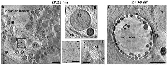
Senior author of this new study – which uses cutting-edge Cryo-Soft X-ray Tomography (Cryo-SXT) techniques to investigate the bacteria – is Dr Maud Dumoux. Dr Dumoux, Technology Lead for Cryo-imaging at the Rosalind Franklin Institute, said: ‘Chlamydiae are unique bacteria, as they grow only inside cells. Most strains form a specialised compartment in which hundreds – sometimes thousands – of bacteria multiply and differentiate themselves between non-infectious and infectious forms, preparing for release into the wider environment to infect other cells in the body.
‘How communities of Chlamydiae organise these events, and how each community has an impact on its individual bacteria, are intriguing questions that that have implications beyond this particular pathogen.’
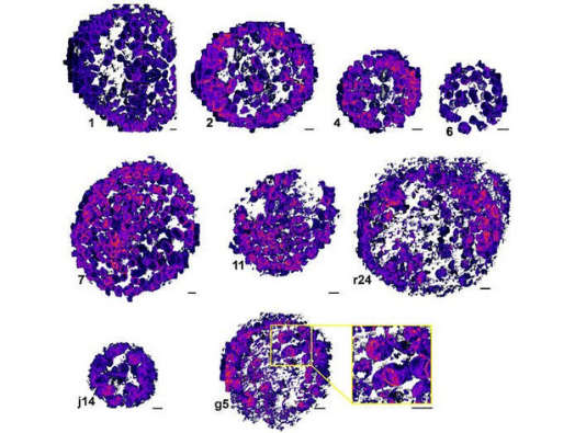
In this paper, the research team – from the Franklin, Diamond Light Source, Research Complex at Harwell, and Oxford University – sought to uncover how communities of Chlamydiae bacteria regulate and organise their space, and how basic factors such as concentration and size of bacteria can impact the life cycle. To do this, the researchers used Diamond’s Cryo-SXT technology – a combination of X-ray tomography and high-resolution fluorescence microscopy that allows scientists to capture intra-cellular behaviour and interactions quickly and in intricate detail.
Dr Dumoux said: ‘Surprisingly, we found that concentration of bacteria is not correlated with differentiation into the infectious form. In other words, at the time we observed the infected cells, a high concentration did not trigger preparation for exiting the cell and infecting other cells.
‘However, we did show that higher concentrations lead to smaller individual bacteria, whereas bacteria given space can reach very large volumes – similar to the apocryphal idea that fish will grow to the size of their pond.
‘This is very interesting because it demonstrates, as is often the case in life science, that things are not black and white. Here, there are shades of grey as the bacteria adapt or respond to their environmental pressures. It also opens up new questions about whether different types of bacteria have different roles within the community.’
The researchers conclude that each ‘inclusion’ (specialised compartment of bacteria) operates as an autonomous community that influences the characteristics of individual bacteria within it – and that bacterial concentration is a key factor in determining those characteristics. With Cryo-SXT now established as a useful and rapid technique for studying Chlamydiae, future research will shed light on the evolution of infection and communication between bacterial communities, which could open up new therapeutic opportunities.