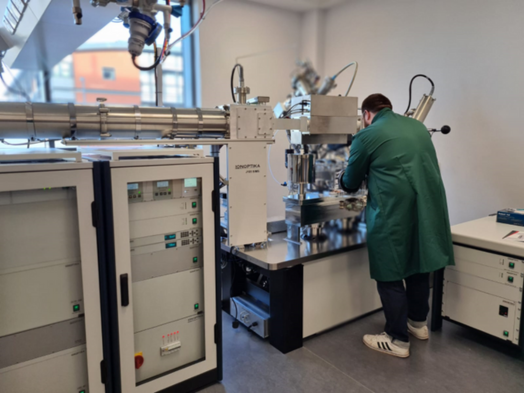
J105 at the Franklin
The J105 SIMS (J105) is a special 3D imaging Time of flight Secondary ion mass spectrometer (ToF SIMS) instrument manufactured by Ionoptika.
SIMS is a technique used to look at the composition of surfaces by hitting the specimen with a focused ion beam and analysing ejected secondary ions to create a 3D image. However, the J105 differs from other ToF SIMS in a number of different ways including a water cluster ion beam and the ability to analyse fragment ions which gives it high sensitivity to biological molecules at a mass accuracy of < 5 ppm.
The J105 was originally installed at the University of Manchester where the Franklin team worked with the Professor Nick Lockyer’s group to develop the instrument and were able to observe large biomolecules and their multiple charge species prior to it being installed in the Franklin. This work in multiomics imaging using water gas cluster ion beam SIMS was published in collaboration with work from partners at Pennsylvania University in Analytical Chemistry.
Prior to its installation, Felicia (who oversees the SIMS project at the Franklin) said about the J105 “We are excited by the arrival of this unique instrument, following on from the work at Manchester we believe it has great scope to enable a broad range of research across multiple Franklin area including imaging of ATP in mitochondria and intracellular imaging of metabolites.”
At the Franklin the J105 is being used for the direct analysis of biomolecules and metabolism in tissue under cryo conditions to study intercellular mechanisms of action in disease. The SIMS team plans to work with groups across the Franklin comparing the images produced by different techniques under cryo conditions such as focussed ion beam scanning electron microscopy (FIB-SEM) and transmission electron microscopy (TEM). Mass spectrometry imaging (MSI) allows specific identification of a broad spectrum of different molecules such as from drugs and lipids, whereas TEM enables structural identification of small regions at the molecular scale (sub-nanometre). SIMS bridges the scale between standard MSI techniques that can resolve tissue structures (at the 10 micron scale), and SEM/TEM imaging, with the SIMS ability to image at the intercellular (sub-micron) scale. The limitation of the SIMS technique is that it fragments the molecule of interest, such that only fragments or small molecules are detectable. The developments in the J105 using a giant water cluster beam have started to push back these limits with the ability to observe intact large biomolecules such as ubiquitin and lysozyme.