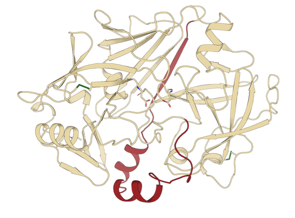
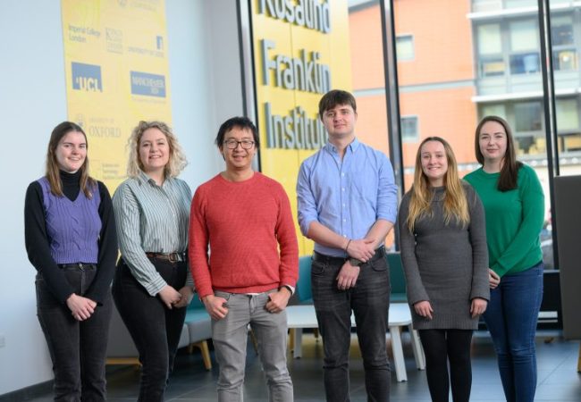
Capturing Complexity: How AntigenApp Is Transforming Nanobody Discovery at the Franklin
Researchers at the Franklin studying schistosomiasis- a neglected tropical disease affecting more than 200 million people globally- have now observed the structure of a key protein thanks to a unique blend of nanobody discovery and advanced data science. Cracking the…

New electron microscopy method opens window into structural studies of thicker, more complex biological samples
Scientists at the Rosalind Franklin Institute have demonstrated a novel way to recover high-resolution structural details of biological objects using transmission electron microscopy – a technique that could reveal new details from thicker, more complex specimens than before. The approach,…
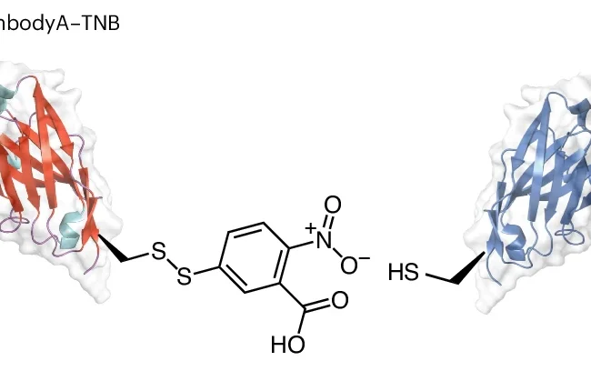
New bifunctional, bispecific nanobodies scaffold helps with small protein imaging
Researchers at the Rosalind Franklin Institute, University of Oxford, and Diamond Light Source have combined their expertise to create new scaffolding molecules to allow the electron microscopy imaging of small proteins (below 50 kDa). Using this method the team have…
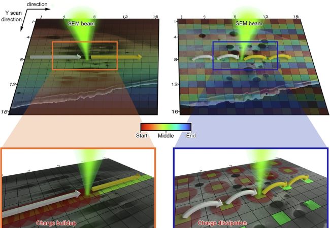
New scanning technique overcomes longstanding challenge in cryo-SEM imaging
Researchers at the Rosalind Franklin Institute have developed a new approach to scanning electron microscopy (SEM) that dramatically reduces obstructions caused by charging artefacts in frozen biological samples. The advance, described in a paper published in Nature Communications, allows scientists…
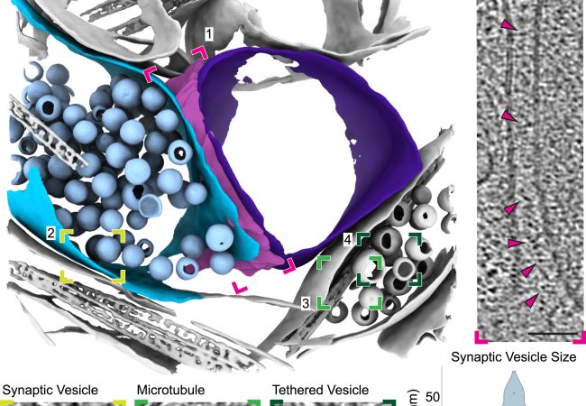
Moving from cells into tissue: a new tomography method
Researchers led by Dr Michael Grange at the Rosalind Franklin Institute have developed a robust molecular imaging workflow for brain tissue samples. Their approach utilises plasma ion beams, which increased the size of samples that could be extracted for imaging,…
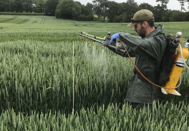
Sugar signalling applications could boost wheat yields by up to 12%
Long term study confirms effectiveness of new technology Enhancing wheat plants ’ sugar signalling ability could deliver increased yields of up to 12%, according to researchers from Rothamsted, Oxford University and the Rosalind Franklin Institute. That is an order of…
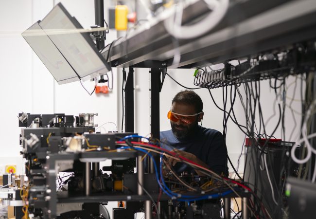
BioCOP – the UK’s first multimodular optical microscope – enters testing phase
Biophotonic Correlative Optical Platform (BioCOP), which is located at the Rosalind Franklin Institute (the Franklin), has entered its next phase of development of calibration and benchmarking. The instrument is now assembled, and the team will start testing it with model…
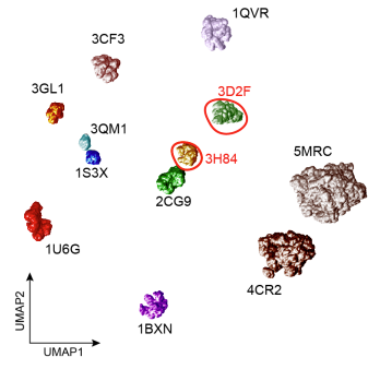
Using AI to improve the analysis of 3D biological images
Scientists have developed a new machine learning model, known as Affinity-VAE, that improves the analysis of 3D biological images in fields such as cryo-electron tomography (cryo-ET). By making use of prior knowledge about protein structures, the model can identify clusters…
Uncovering the hidden lives of complex sugars with cutting-edge imaging
Dr Liang Wu is a Wellcome Trust Sir Henry Dale Fellow at the Rosalind Franklin Institute. Since joining the Franklin in 2020, Dr Wu’s team has been investigating the role and function of enzymes in the production of heparan sulfates…
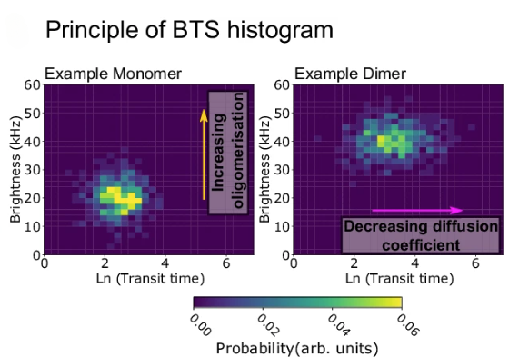
New technique sheds light on how proteins organise and move on cell membranes
Researchers at the Rosalind Franklin Institute, the University of Oxford and the University of Southern California have developed a new method for studying how molecules behave on the cell membrane. The technique, known as brightness-transit statistics (BTS), will enhance our…