Dr Judy Kim

Judy Kim is the Deputy Challenge Lead for Multidimensional imaging at the Rosalind Franklin Institute and Departmental Lecturer in the Department of Materials at the University of Oxford.
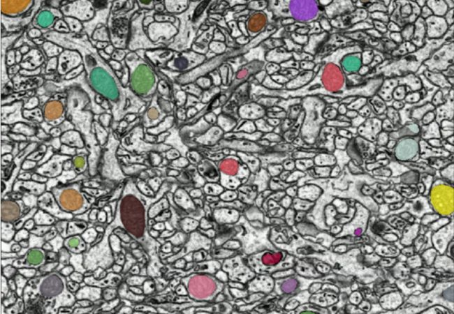
Her work brings together the new developments in transmission electron microscopy with practical application in beam-sensitive biological materials. Judy’s work with the Multidimensional Imaging team pushes the boundaries of what is possible using:
- Defocused-probe ptychographic imaging for high-contrast, high-resolution, large field of view scans of nanostructures internalised in cells
- Microsecond resolution in situ TEM videos of membrane dynamics in liquid flow cells
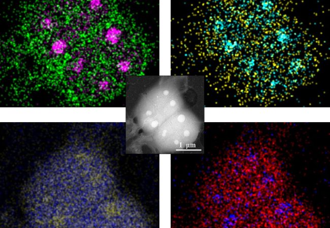
- Electron X-ray Dispersive Spectroscopy in conjunction with scanning techniques for elemental maps of markers in cells or on chromosomes
Judy has a special interest in the various forms of in situ TEM and pushing the current boundaries of temporal resolution using novel optical setups, modified electron sources and modern detectors for application towards radiation sensitive materials.
Her previous projects at the University of Oxford, focused on technique development using monochromation and aberration correction for characterization of energy and 2D materials. Her PhD work at the University of California, Davis contributed to the development of a Dynamic Transmission Electron Microscope where she was awarded a Lawrence Scholar Program Fellowship from Lawrence Livermore National Laboratory. Her study on exothermic nanolaminate reactions led to the first publication of nanosecond + nanometer scale images and was awarded the Zuhair Munir Best Dissertation Award. She has also received the R&D 100 Award in 2008 and the Microscopy Today Innovation Award 2010 for her development work on the DTEM.
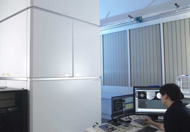
Aberration-corrected transmission electron microscope
Ruska is an aberration-corrected transmission electron microscope (TEM) used to explore novel methods to study radiation sensitive specimens such as biological materials that have been cryogenically preserved or encapsulated in liquid for dynamic observations.
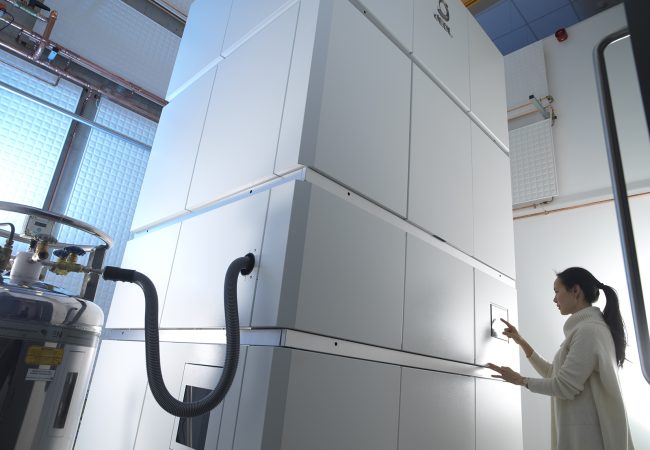
Chromatic Correction
Knoll, the first chromatic aberration-corrected electron microscope in the UK housed at the Franklin, will push the current resolution limits for biological samples by correcting energy variations in the electron beam.
Cryo-ptycho-tomography
Developing a novel technique using cryo-electron ptychography to perform tomographic characterisation of biological processes at cellular scales, enabling detailed study of rare and complex structures in their native environments.
Liquid Phase Electron Microscopy and Spectroscopy
Transient, dynamic assemblies of biomolecules in solution are the primary driving forces behind biology. However, studying these at high resolutions requires the use of electron microscopes (EM), which need extremely high vacuums to function.
Citizen science
People-powered research, or citizen science, is the participation of non-experts in scientific research. There are many ways to take part in citizen science, but one popular way is through the Zooniverse platform.
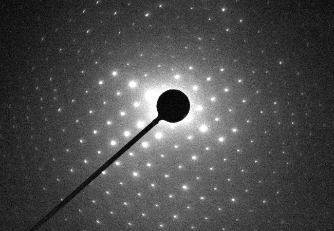
Electron diffraction
MicroED is an emerging technology that exploits the strong interaction of electrons to reveal the structures of molecules from vanishingly small crystals.