Researchers measure small forces inside immune cells using advanced microscopy
Scientists from the Rosalind Franklin Institute and the Kennedy Institute at the University of Oxford have, for the first time, measured the small mechanical forces generated by cells during an immune response.
Using a combination of advanced microscopy techniques, the researchers have developed a method of measuring the mechanical forces in 2D and 3D at unprecedented sensitivity.
The research is published in two new papers in Nature Communications and paves the way for greater biomechanical understanding of how our immune cells respond to harmful pathogens.
Senior author Professor Marco Fritzsche leads the Biophysical Immunology Laboratory – a joint venture between the Rosalind Franklin Institute and Oxford’s Kennedy Institute for Rheumatology. He said: ‘Physics plays a profound role in a range of biological processes in the human body. One of these scenarios is the protective function of our immune cells, which fight off disease by binding to the antigens being carried by infectious agents.
‘Each of these molecular interactions involves the generation of small mechanical forces that help immune cells differentiate between antigens from their host body or from invasive pathogens. Until now, we haven’t been able to measure those forces at the right sensitivity.’
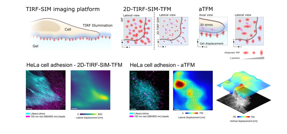
The researchers developed this new force-probing technique by combining astigmatic traction force microscopy and TIRF-SIM microscopy with traction force microscopy to improve the overall spatial and temporal performance of the methodology.
Professor Fritzsche added: ‘This 100-fold improvement in temporal resolution provides enhancements equivalent to mechanical forces on the nano and sub-second scales.
‘The establishment of this technique has the potential to reshape biomedical science in years to come. Now, for the first time, we can measure the mechanical forces generated by cells during an immune response. We can begin to understand the influence of the smallest mechanical forces in health and disease, and ask why cells can be so successful in recognising foreign antigens under some conditions but not, in the case of autoimmune diseases or cancers, in others.’
The work was led by Dr Huw Colin-York and DPhil student Liliana Barbieri, both members of Professor Fritzsche’s group. It was carried out in close collaboration with Professor Michael Dustin at the Kennedy Institute, Professor Dong Li at the Biophysics Institute at Beijing, and Professor Emad Moendarbary at UCL.
A discussion piece on these novel microscopy techniques has also been highlighted by the Royal Society: