Chromatic Correction for observing biological dynamics
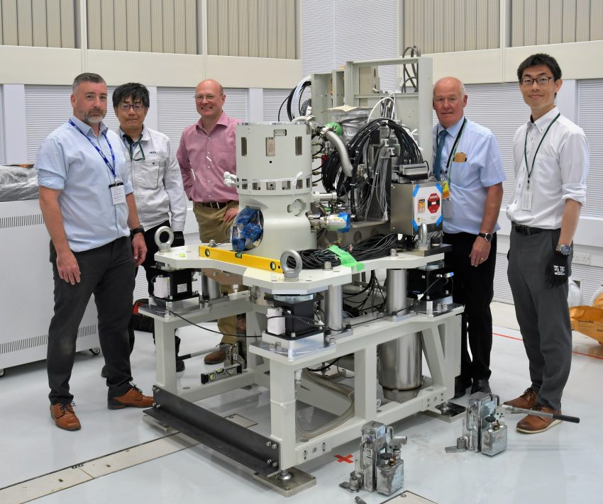
JEOL’s pioneering chromatic aberration corrected electron microscope has arrived at the Franklin. This is the culmination of a five-year collaboration which was started at the outset of the Franklin and has already delivered two other unique instruments. The development of the latest instrument represents the first of its type globally and the first chromatically corrected instrument in the UK. Commissioning will take place over the next year by a team at the Franklin that includes a JEOL embedded research scientist for 3 years. Initial applications of the instrument will be to use sophisticated liquid cells to study biological dynamics and to study thick samples.

Background
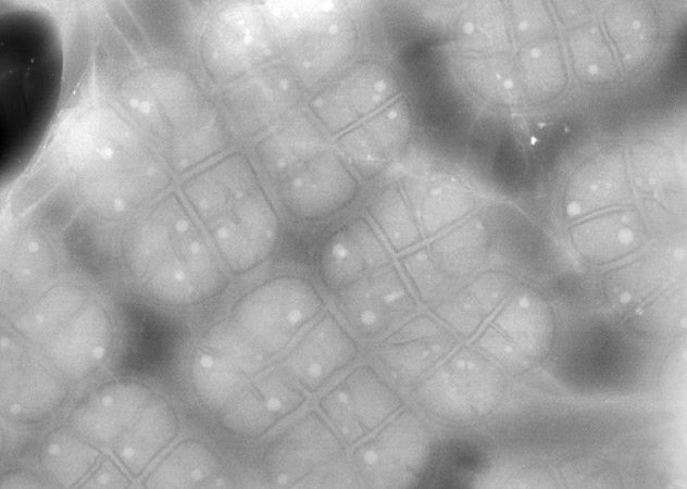
To be able to observe molecular dynamics and interactions it is essential that the biological sample is in a liquid state. Liquid cells for electron microscopy consist of two membranes that seal a film of liquid from the microscope high vacuum and as such are considerably thicker than conventional samples used for electron microscopy. This leads to a loss of resolution due to chromatic blur arising variable focussing of inelastically scattered electrons limiting high resolution dynamic imaging. To overcome this, we have co-developed a novel electron optical chromatic aberration corrector to overcome limitations in spatial resolution and which has been designed to allow high electron flux to be used; an essential component to achieving high temporal resolution. This complex optical element is now installed in a new state of the art column at the Franklin optimised for studies of thick samples.
Science in detail
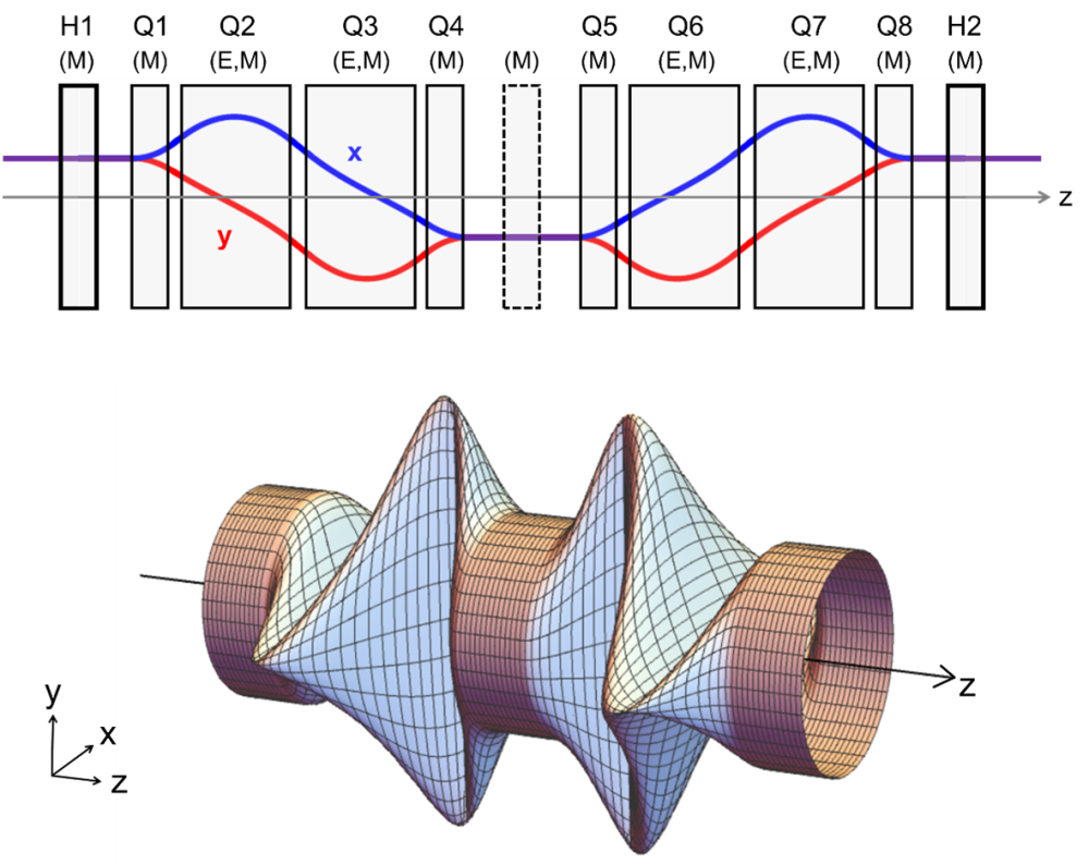
All electromagnetic round objective lenses suffer from inherent spherical and chromatic aberration which cannot be overcome by improvements in mechanical or electrical engineering. The only practical solution to this is to introduce non-round elements which to correct chromatic aberrations requires a combination of magnetic and electrostatic fields to form a Wien condition.
Working with JEOL we have developed a chromatic and spherical aberration corrector based on a combination of hexapole and quadrupole fields. Thick hexapole fields are used to generate negative spherical aberration and to correct residual axial and off-axial aberrations. However, instead of using round transfer lenses placed between the hexapoles, a quadrupole multiplet producing superimposed electric and magnetic quadrupole fields is used to produce negative chromatic aberration . The quadrupole multiplet also functions as a transfer doublet within the corrector. This latter feature eliminates point foci enabling higher flux densities to be transferred without space charge energy broadening, which is essential for fast dynamic studies that are limited by the electron flux.
Impact
The new instrument including the corrector has arrived at the Franklin and is currently being commissioned. As part of the collaboration with JEOL we have a research scientist who was the group lead for the development from JEOL based with us full time to work on this project. Our initial experiments will include the use of novel in-house developed graphene cells for studies of dynamics of small molecules. More conventional liquid cells where we can change the local liquid environment will be used to study the dynamics of environmental and chemical stress, and pharmaceutical uptake. The new instrument will also advance tomography where samples at high tilt become too thick for high resolution imaging.
Why the Franklin?
This research was only achievable at the Franklin for a number of reasons; firstly, we established an early long term collaboration with JEOL based on basic research; secondly the instruments delivered which have been co-developed have provided a research platform for new methods that have already been used in a number of applications.