
New milestone reached with unique electron microscope for tomography: years in the making and brought to life through close collaboration.
The Rosalind Franklin Institute and Thermo Fisher Scientific have reached a major milestone in their long running collaboration to develop a unique research instrument based on the Krios cryo transmission electron microscope. Specially configured to address some of the challenges in imaging cells and tissues, the system has now completed installation, commissioning and acceptance and is ready to move into its next phase: enabling new science.
Getting here has taken an extraordinary collective effort.
Scientists, engineers and technical teams across the Franklin and Thermo Fisher Scientific have worked closely together over several years, taking the project from an ambitious scientific idea through technology development and engineering to installation, testing and optimisation. The timelapse captures only the final stages of a much longer journey involving many people across both organisations.
The scientific ambition behind that effort is significant.
“We’re incredibly proud to be at the forefront of cryo-EM, and excited by what this partnership with the Franklin has made possible. By bringing our teams’ expertise together, we’ve turned an ambitious scientific challenge into a new capability for researchers.”
This unique instrument has several new developments with improvements made with improving electron tomography in mind and to allow the researchers to gain the most insight into every important biological sample it images. This instrument utilises a specialised optical component allowing for chromatic aberration correction, which allows the researchers to capture information from electrons that would be lost with a standard electron microscope set-up.
Cryo electron microscopy allows biological samples to be preserved at very low temperatures so researchers can examine the molecular machinery of life in extraordinary detail. However, as samples become thicker and more representative of the complexity of a cell, they need to be exposed to the beam of electrons for longer doing so increases the damages to the sample.
To potentially extend the electron budget to allow for greater signal to noise, the microscope has been adapted to operate at lower temperatures. Whilst typical cryogenically cooled transmission electron microscopes operate around 77K or -180 degrees Celsius (liquid nitrogen temperature), this tool has a novel battery cooler allowing the instrument to operate at 25 Kelvin or -247 degrees Celsius.
During his recent visit to the Franklin, Science Minister Chris McDonald MP had the opportunity to see the instrument and meet some of the people behind the programme, hearing first-hand about the research possibilities this new capability could enable.
Dr Michael Grange, Emerging Interest Area Lead for Structural Cell Pathology, has been involved in the development of this instrument, said: “We are excited to get started characterising this unique instrument. My team plan to utilise these advances to look for some of the first molecular changes that happen in the onset of brain diseases, which will hopefully unlock new knowledge which could lead to the development of new treatments.”
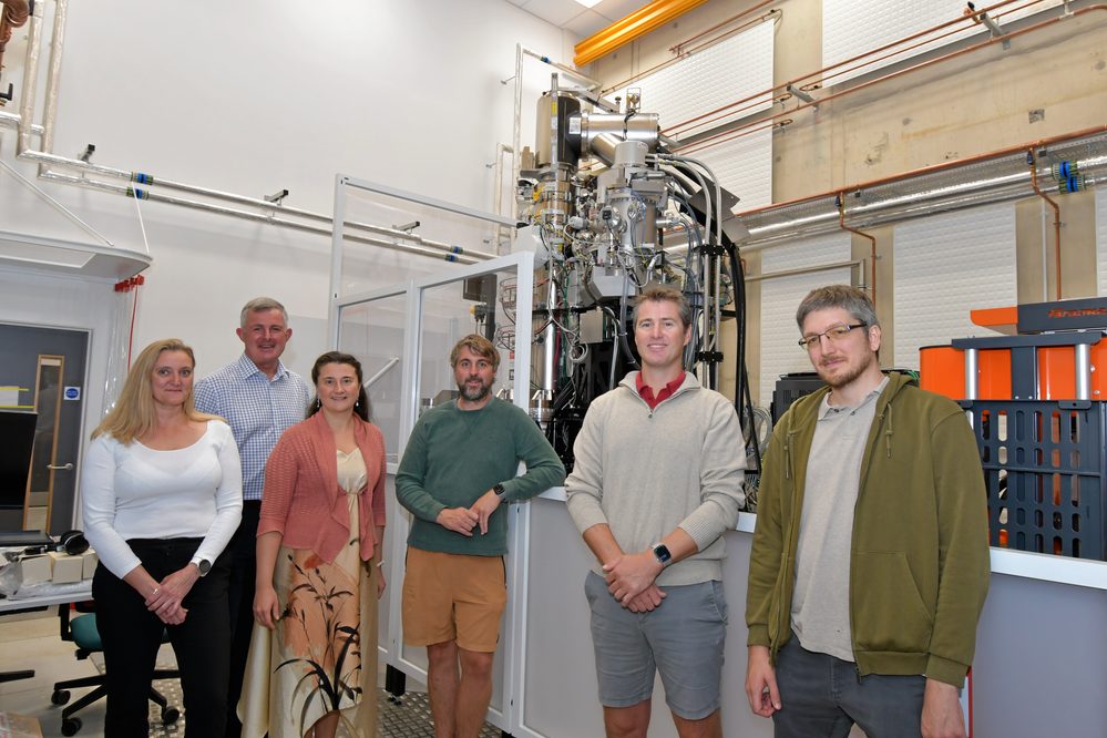
The research team will be imaging surgical brain biopsies from patients to try to understand how changes in structures in the brain may change during aging or the onset of disease. The aim is to try to understand early disease changes that may impact our fundamental understanding neurodegenerative disease.
The team at the Franklin have been focused on optimising sample preparation and automation of this process over the past few years. Now the emphasis changes – to see what new resolution can be reached and looking at the wider molecular context in biological samples.
In addition to applying this new technology to study neurodegeneration, the Franklin team are keen to work with the community and provide training on these cutting edge techniques.
Dr Maud Dumoux, Deputy Challenge Lead for Quantitative Biology Across Scales, said: “What makes this project particularly exciting is that it was driven by a scientific challenge that could not be addressed with existing instrumentation. Working closely with Thermo Fisher Scientific has allowed us to develop and integrate new technologies specifically aimed at extracting more information from precious and complex biological samples.”
“This is not only about building a unique microscope, but about making the new science it enables. We want to work closely with the community, develop applications together and train researchers so that these approaches can be adopted much more widely.”
“This is not only about building a unique microscope, but about making the new science it enables. We want to work closely with the community, develop applications together and train researchers so that these approaches can be adopted much more widely.”
A huge thank you to the many people across Thermo Fisher Scientific, who have helped make this possible. We are excited to see where the science takes us next.