This work will improve understanding of long-term infection and could guide new ways to study and treat similar hard-to-target intracellular bacteria.
Understanding how Chlamydia adapts to life inside and outside host cells
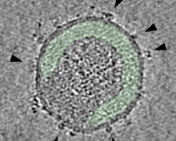
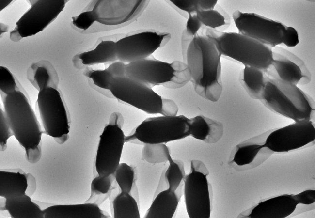
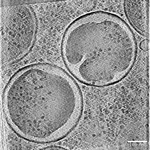
We are studying how Chlamydia changes form to infect and survive in the body. Using advanced imaging and protein analysis, we track how its internal machinery adapts.
Technical detail
Chlamydia is a common sexually transmitted bacterial infection with an estimated 128.5 million new infections occurring worldwide in 2020 (WHO). Chlamydia infection is often asymptomatic but can cause serious health problems if untreated, such as infertility and increased risk of other infections.
Chlamydia is an obligate intracellular pathogen that transitions between distinct developmental forms to infect, survive and persist within its host. Despite its medical importance, the molecular mechanisms that drive these transitions remain poorly understood because the organism is difficult to genetically manipulate and study using conventional approaches.
At the Rosalind Franklin Institute, we are combining state-of-the-art cryo-electron microscopy, structural biology, and time-resolved proteomics to investigate how key molecular machines, including the Type III Secretion System (T3SS) and bacterial ribosomes, are remodelled during the Chlamydia life cycle.
By correlating protein production with high-resolution in situ structures, we aim to reveal how the bacterium adapts between its infectious elementary body, replicative reticulate body, and persistent stress-resistant forms. This unique multidisciplinary approach has the potential to uncover fundamental mechanisms of host–pathogen interaction, provide new insights into chronic infection and persistence, and establish a broadly applicable framework for studying other intracellular pathogens.
Project Team

Collaborators


Contact information

PhD Studentships
Applications now open
Related projects
-
![]()
Visualising HIV entry one virus at a time
-
![]()
Applying artificial intelligence to accelerate the discovery of anti-viral nanobodies
-
![]()
Experimental automation
-
![]()
Defining how coronaviruses enter and traffic in cells
-
![]()
Liquid Phase Electron Microscopy and Spectroscopy
-
![]()
Electron detector development
-
![]()
Electron diffraction
-
![]()
Cryo-ptycho-tomography