This work helps us understand how HIV infects cells and could reveal new targets for vaccines and antiviral treatments.
Visualising HIV entry one virus at a time
This project explores how HIV enters human cells by studying proteins on the virus surface and how they work together. Using advanced imaging, we observe how these proteins move and cluster.
Technical detail
HIV (human immunodeficiency virus) is a virus that attacks the immune system, weakening the body’s ability to fight infections and disease, specifically affecting white blood cells. There were 1.5 million HIV infections in 2020 (WHO).
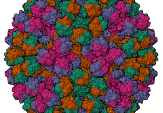
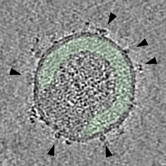
HIV infection begins when the viral envelope protein (Env) engages receptors on the surface of immune cells and triggers membrane fusion. While the structure of individual Env proteins has been studied extensively, much less is known about how multiple Env molecules are organised and behave on the surface of intact viruses, and how this organisation influences viral infectivity and immune evasion.
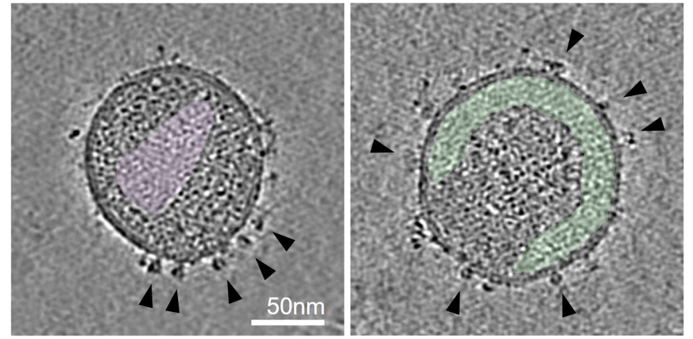
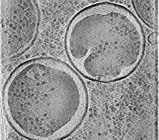
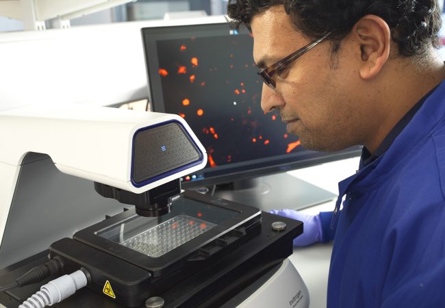
This project combines advanced fluorescence microscopy, cryogenic imaging, single-molecule tracking and cryo-electron tomography to visualise HIV particles in unprecedented detail. By studying how Env proteins cluster, move and respond to host factors that naturally restrict infection, we aim to uncover the molecular mechanisms that govern viral entry into cells. The work brings together expertise in virology, biophysics, structural biology and correlative imaging from King’s College London, Biofisika Institute, University College London and the Rosalind Franklin Institute, with the goal of revealing new vulnerabilities in HIV that could be exploited for future antiviral therapies and vaccine design.
Project Team

Collaborators


Contact information

PhD Studentships
Find out more here