Advanced Methods and Algorithms for Electron Ptychography of Biological Specimens
Electron ptychography is a powerful, dose-efficient imaging technique for studying biological specimens. This project will develop improved scanning and phase recovery methods to reduce the electron dose needed even further. This could enable high-resolution imaging of more delicate samples and reveal biological structures that are currently difficult to study.

Electron ptychography is an advanced imaging technique that uses 4D scanning transmission electron microscopy data to produce high-resolution images of biological specimens. However, electron beams can damage biological samples, limiting the quality of information that can be collected.
This project will develop improved scanning and reconstruction methods to obtain clearer images using less electron dose. The initial focus will be on optimising how the microscope scans a sample and how the resulting data are processed, while accounting for practical factors such as image quality, acquisition time, sample movement and instrument stability.
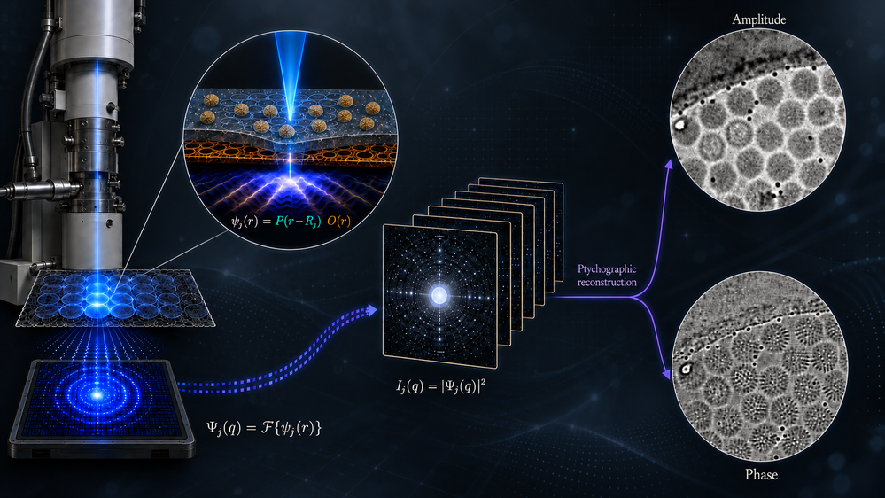
The research will combine mathematical modelling, algorithm development and validation using experimental electron microscopy data. The methods will be designed to work with Franklin’s existing instruments and established sample-preparation workflows, without requiring major hardware changes.
By making electron ptychography more dose-efficient and robust to common experimental imperfections, the project aims to support high-resolution imaging of fragile biological samples and enable deeper understanding of biology.